Patterns of Sleep Disorder in Alzheimer’s Dementia- A Scoping Review
Ayobami Yusuff and Ioana Toma
Abstract
Introduction
Alzheimer’s disease (AD) is the most common type of dementia, characterised by progressive cognitive deterioration alongside other symptoms. Amongst the non-cognitive symptoms, 25 to 40 % of patients suffer from disturbed sleep patterns. Sleep is essential to the brain as it supports learning and memory, regulates synaptic plasticity, and enhances waste clearance.
Method
This review was set out to explore and understand the possible patterns of sleep disorder in AD, adding to the existing knowledge about this condition of growing health significance.
This is a systematic search of the literature in the EBSCO host databases. The keywords for the search criteria in the Boolean operators of multiple databases were words that describe ‘Alzheimer AND sleep disorder’, done up to 9 January 2024.
Results
A total of 1,511 records were obtained, 118 were read in full text and only 20 of these met the study criteria. The results demonstrated macroarchitectural sleep changes, which were 45% of the total results. Other results included reduced sleep drive, broad REM sleep changes and reversal of normal physiological sleep pattern; microarchitectural sleep changes are higher N1 and lower N3, reduced alpha activity during wakefulness and prominent EEG slowing in REM sleep.
Discussion
There are diverse sleep changes in AD which impact its presentation and course. Sleep disorders may also predate cognitive decline and may therefore be a potential biomarker for AD and may be used to discriminate the stages of its severity. We recommend that sleep history should be incorporated into the holistic assessment and management plans of patients with AD.
Introduction
AD accounts for most types of dementia cases and is characterised by insidious progressive cognitive deterioration alongside a constellation of other non-cognitive symptoms [1]. At the molecular level, it is a progressive neurodegenerative disorder characterised neuropathologically by the accumulation of amyloid-β as extracellular plaques and intracellular neurofibrillary tangles of hyperphosphorylated tau [2]. Cognitive symptoms include short-term memory dysfunction, executive dysfunction, reading and calculation impairment, comprehension and judgement difficulties, and attentional struggle. Within the category of non-cognitive symptoms, we find mood disturbances, psychotic symptoms, behavioural alterations such as agitation, aggression, wandering and pacing, and changed sleep patterns [3].
Disordered sleep patterns in AD
About 25% to 40% of AD patients suffer from disturbances of sleep, such as insomnia, excessive daytime sleepiness, rapid eye movement (REM) sleep behaviour disorder, sleep apnoea and circadian rhythm disturbance [3]. Another type of agitated behaviour in AD is sundowning, which is described as the occurrence or exacerbation of behavioural symptoms of AD in the afternoon and evening, closely related to disturbance of the circadian rhythm [4]. Sleep problems occur early in the course of AD, and disordered sleep has been associated with an increased risk of developing AD [5].
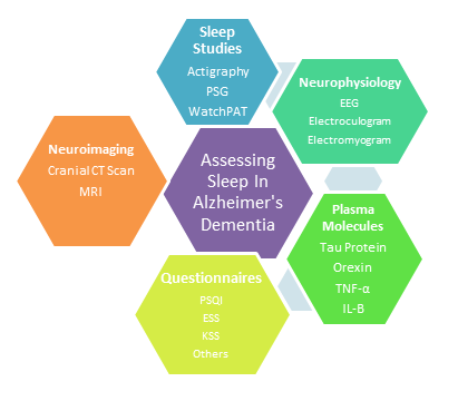
When assessing sleep disorders in AD, several methods have been applied.
Figure 1: Subjective and objective assessments of sleep in patient populations. Image adapted from Winsky-Sommerer et al [6].

Epidemiology of Sleep Disorders in AD
Disturbed sleep is one of the most reported symptoms in people with dementia [7], but estimates of its prevalence vary significantly, ranging between 5 % and 86 % [8]. Differences in the measures used to identify the sleep problems, dementia subtypes, and dementia severity of people in the sample being studied might all contribute to this variability in estimates.
The prevalence of sleep disturbance may vary depending on the setting in which the persons with dementia live, as community-dwelling individuals and those living in long-term residential settings are subject to numerous environmental differences, such as privacy, noise levels and the presence of specialised carers versus family caregivers. As a result, it is crucial to differentiate these two population groups when analysing sleep disturbance patterns, as around 84% of people with dementia worldwide live in the community [9]. Moreover, sleep disturbances are a predictor of people with dementia living in the community moving into care homes [10].
The multifaceted nature of geriatric sleep impairment is believed to reflect a complex interaction between a variety of factors, including age-dependent decrease in the ability to sleep, neuropsychiatric comorbid disorders such as depression and dementia, pain and limitation of mobility with excessive bed-time, increased prevalence of sleep-disordered breathing and nocturnal myoclonic activity, and disturbed behaviour during sleep including nocturnal confusion (sundowning) [11].
Accumulating evidence shows that sleep disturbance contributes to cognitive decline and might also increase the risk of AD [11]. Thus, it would be helpful to understand the patterns of sleep disorder (SD), as this can potentially guide clinicians in designing care plans and incorporate and address carer burden. Furthermore, emerging research has provided a direct link between several of these sleep disruptions and AD pathophysiology, suggesting that treating sleep disorders in this population may target basic mechanisms of the disease [12].
Aim
There is growing evidence that sleep disorder in AD is a core part of clinical presentation; however, little is known about the characteristics and manifestations of SD in AD. This review aims to identify and summarise current patterns and characteristics of sleep disorders in AD.
To the best of our knowledge, this is the first article synthesising multiple reviews that describe the patterns of presentation of sleep disorder in AD. To understand the implications of the potential relationship between the two conditions, we formulated the following research questions:
What are the relationships between sleep disorder and AD?
What are the patterns and characteristics of sleep disorder in AD?
What are the potential clinical indications for identifying them?
Method
Using the methodology of a systematic review but adopting a scoping review, we were able to qualitatively extract and describe the results. The Preferred Reporting Items for Systematic reviews and Meta Analyses (PRISMA) extension for scoping reviews was used as a guideline in writing this project. On the hierarchy of evidence, all studies with a level of III or above were considered. We used the Patient/Population, Intervention, Comparison, and Outcomes (PICO) framework to appraise each study.
A systematic search of literature in EBSCO host Research Databases, including: EMBASE, PubMed, MEDLINE, CINAHL, and GreenFILE was conducted until 9 January 2024; there was no limit to the starting search date. The keywords for the search criteria in the Boolean operators were words that describe ‘Alzheimer AND sleep disorder’. The following keywords were used: neurodegeneration and sleep disturbance, neurodegeneration and sleep disorder, Alzheimer and sleep disturbance, dementia and sleep disorder, and dementia and sleep disturbance.
The study criteria used were: studies published in the English Language and performed on human subjects, not animal models, not AI-generated reports and clearly answer the aims of the literature review, i.e., focus on describing the patterns of sleep disorder in AD.
After an initial stage of reading the title and abstract of each study generated from the search, studies that met the research aim were extracted. The first grade of collected studies were then read in full text to identify those that specifically answer the research question.
We invited an independent reviewer to inspect the search criteria, the findings, all abstracts and then the full texts identified from the searches. The process was facilitated by the use of EBSCO host folder sharing.
As a literature review, it is generally considered low-risk research with no direct contact with human subjects and so no specific ethics application was sought; however, for each article referenced in this project, appropriate ethics have been considered as described by the authors.
1,511 results were obtained in the initial search. 219 duplicate articles were automatically removed. 118 publications were read thoroughly and only 20 were included in this review.
Figure 2: Flowchart of the literature review

Results
Figure 3: Methodologies identified
We classified the disturbed sleep patterns broadly and vaguely into macroarchitectural changes and microarchitectural changes, though this may not be applicable in all the results of this review, as some articles correlated sleep disturbance with other parameters such as the proteinopathies and genetics of AD.
Macroarchitectural sleep changes
Table 1: P-Patients, C-Controls
| Authors and year |
Sample size
|
Method/ Parameters measured | Findings/Results | Values |
| Devulder et al 2023 |
P- 41 C-30 |
24h EEG PSG |
Reduced REM Increased WASO Reduced SE Longer TIB
|
REM reduced by <6%; REM < 33 mins WASO > 72 mins; P=.001 SE reduced by < 12%, P=.087 |
| Liguori et al 2016 |
P- 20 C- 26 |
EEG PSG MRI CSF Orexin
|
Negative correlation between CSF Orexin and REM | R = −0.65, p = 0.001 |
| Vitiello et al 1990 |
P- 44 C- 45 |
PSG |
WASO significantly greater in the AD group AD subjects spent significantly more TIB and had significantly less %3-4 TIB REM latency measures were significantly shorter in the AD group
|
P=.006
P=.03
P=.04; p=.01 |
|
Tadokoro et al 2020
|
P= 41 C= 22
|
PSQI WatchPAT
|
Bad self-check PSQI scores in AD vs NC
Decreased REM sleep in AD
Increased NREM sleep in AD |
PSQI>5 (42.9%) vs (81.8%, * p < 0.05)
P<0.05
P<0.05 |
| Gagnon et al 2006 |
P= 15 C=15 |
EEG Infrared video |
Shorter sleep time in AD Lower nr of REM periods in AD Higher percentage of stage 1 sleep in AD RSWA in Ads
|
REM sleep and Sleep Efficiency (SE)
REM sleep changes and reduced SE have been consistently reported [13,14,15,16]. AD patients have been observed to have less REM sleep plus shorter REM latency than their healthy counterparts and this has even been observed at the stages of subjective cognitive impairment, prior to the clinical assessment or diagnosis of AD [15]. While REM sleep is generally associated with low muscle tone, a small number of patients with AD have abnormal motor activity in REM sleep, a significant number of them do have REM sleep without atonia (RSWA) [14].
Wake after sleep onset (WASO) has also been reported in AD and has been identified as a predictor of progression of mild cognitive impairment (MCI) to AD [13,16]. Sleep efficiency (SE) is described as a ratio of the time spent asleep to the total time in bed (TIB). A sleep efficiency of 85 % or higher is normal, while a sleep efficiency anywhere above 90 % is considered very good. While reporting SE, Alzheimer’s sufferers spent shorter sleep time [14], more time awake, plus high number of nocturnal awakenings, and more TIB [17]. Overall, there is reduced SE in AD.
Some reports show slightly different results; for example, WatchPAT analysis showed REM sleep decreased significantly in AD and slightly in MCI compared with normal controls (NC) [18]. Non-REM light sleep increased significantly in AD and slightly in MCI compared with NC, but not deep sleep. These findings also demonstrated occasional differences between objective and subjective reports/assessments of sleep in AD, although objective assessments are still preferred options for clinicians.
Disturbance of physiological sleep timings
Table 2: P- Patients, C- Controls
| Authors and year | Sample size | Method/ Parameters measured | Findings/Results | Values |
| Tractenberg, E., Singer, M., and Kaye, A. (2006) |
263 AD 399 controls |
SDSQ IADL Cornell Depression Scale |
SP had higher SDSQ scores and more no of symptoms People with SP had worse depression scores
Six symptoms were more frequent in AD + SP: number of wakeups per night; drowsy during day; daytime naps; equal day, night sleep time; trouble breathing; breathes irregularly |
P<.001
P<.01
In both cohorts SL >30 mins
|
| Ancoli-Israel et al (1994) |
246 AD 94 controls |
Sleep questionnaire (10) BDRS DRS POD |
The more demented the patient, the more time they spent in bed, the more fragmented their sleep and more daytime naps they took. Correlation between prolonged sleep, disruptive behaviour and severity of dementing process |
|
| McCurry et al. (1999) | 205 AD |
BDRS RMBPC |
Most frequent sleep problems reported by caregivers was patient sleeping more than usual (40%) and patient awakening early (31%), which caused relatively little distress Other behaviours (e.g., waking the caregiver up at night) were much more distressing. |
|
| Liguori et al 2014 |
P- 48 C- 29 |
PSG CSF Orexin CSF tau and Aβ1-42 |
Earlier bedtimes in severe AD Increased CSF orexin levels were correlated with decreased sleep efficiency, N3 and REM sleep. |
P=.04, r = −0.38; P = .007, r = −0.32 P = .027, r = −0.31 P = .03 respectively. |
|
Mulin et al 2011
|
103 AD
|
Actigraphy
|
Patients with AD and apathy have higher WASO, more TIB and mean duration of daytime inactivity periods
|
P<0.01 P<05 P<0.001 |
|
Park et al 2011
|
36 AD |
UPDRS ESS, KSS PSQI MSLT PSG
|
Patients with AD+PF are more sleepy than those without PF
|
Several reports have described a change in sleep timing, sleep scheduling and reversal of normal day-night sleep patterns. AD patients present with nocturnal awakening [16], earlier bedtimes with daytime sleepiness [19], night-time wakefulness and nocturnal wandering [17], higher duration of daytime inactive periods [20], and a worse daytime dysfunction [21].
In a retrospective cohort study, Tractenberg et al [22] analysed 21 sleep disturbance symptoms using The Sleep Disturbance Symptom Questionnaire (SDSQ), Instrumental Activities of Daily Living (IADL) questionnaire and The Cornell Depression Scale. For less than half of the symptoms in people with AD and a quarter of those in non-demented elderly (NDE), endorsement rates were significantly different depending on whether the reporter (or their sleep partner) did or did not report a sleep problem. In both the cohorts, people with sleep problems (SP+) had significantly higher SDSQ total scores and numbers of symptoms endorsed, and significantly worse depression scores. In both cohorts, SP+ reflected long sleep latencies (>30 min) and a tendency to lie awake worrying, as well as restless sleep, waking too early, waking during the night for at least 1 hour and the use of medication to get to sleep; having bladder and bowel problems at night also followed this trend. The authors suggested that daytime sleep is the most distinctive feature of sleep disturbance in AD patients.
It has been observed that the more demented the patient, the more time they spend in bed, the more fragmented their sleep is, and the more daytime naps they took. There was a correlation between lengthy sleep and disruptive behaviour [23]. Here, it was demonstrated that prolonged sleep is associated with a severe dementing process and behavioural disturbance.
Cluster analysis results revealed three distinct patterns of patient behaviour that were associated with patients awakening their caregivers at night:
(1) mildly demented patients who were fearful, fidgety, and occasionally sad;
(2) patients with few mood or behaviour problems but who spent considerable periods of time sitting alone during the day; and
(3) more severely demented patients with multiple cognitive, depressive, and disruptive behaviour problems [24].
The most frequent sleep problems reported (e.g., sleeping more than usual) caused relatively little caregiver distress, while other behaviours (e.g., waking the caregiver up at night) were much more distressing.
Molecular tests and other Alzheimer’s proteinopathies
Table 3: P- Patients, C- Controls
| Authors and year | Sample size | Method/ Parameters measured | Findings/Results | Values |
| Oh et al 2022 |
P- 53 C- 32 |
EEG and PSG Postmortem immunochemistry |
Greater loss of wake-promoting neurons (LC, LHA & TMN) in AD |
Shorter sleep time- LHA: r = −0.63; P = .009; TMN: r = −0.62; P = .008 Greater WASO- LHA: r = 0.85; P < .001; TMN: r = 0.78; P < .001 Greater number of LC neurons = shorter total sleep time (r = −0.68; P = .008) and greater REM latency (r = 0.71; P = .006). |
| Cooke et al 2006 |
P- 76 C-12 |
PSG; effect of AChEI |
Lower stage 1 sleep in Donepezil vs Galantamine Higher stage 2 sleep in Donepezil vs controls |
P=0.01
P= 0.04 |
| Chen et al 2012 |
P- 43 C- 22 |
PSG ESS |
SE and WASO higher in AD | |
| Durães et al 2023 |
P-78 C-70 |
PSQI CSF Biomarkers |
Sleep duration score was lower in AD Daytime dysfunction worse in AD Negative weak correlations between Ab42 values and daytime dysfunction Weak correlation between t-tau values and daytime dysfunction |
R= – 0.21, P=0.011
R= – 0.16, P=0.046 |
Several Alzheimer’s disease proteinopathies have been associated and correlated with sleep disturbance. In a recent discovery of the glymphatic system- the lymphatic system of the CNS – disturbance of the glymphatic system is mediated through aggregation, impaired clearance, and toxicity of AD proteinopathies (Aβ, APOE4, tau, and others). This leads to a cascade of hypoxia-ischemia and atrophy of the sleep regions in the brain [25]. Although the AD biomarkers were higher in AD patients than controls, there were no statistically significant differences in most studies [16,19].
Orexin system dysregulation may contribute to sleep impairment in MCI patients with a high likelihood of AD pathology, mainly affecting REM sleep and resulting in insomnia, prolonged sleep latency, and nocturnal awakenings [16]. Increased Orexin-A in CSF is associated with sleep fragmentation and sleep disruption. Higher levels of Orexin have been associated with decreased SE and decreased REM sleep [19].
These results imply that in AD, increased CSF orexin levels are linked to a parallel sleep deterioration, which further appears to be related to cognitive decline. Hence, in AD, orexinergic output and function seem to be overexpressed with disease progression and severity, possibly owing to an imbalance in the neurotransmitter networks regulating the wake-sleep cycle toward the orexinergic system promoting wakefulness.
In another report, AD patients carrying the -31TT genotype in the IL-1β promoter plus the APOEε4 allele were susceptible to sleep disturbance [26]. This group showed less TIB, longer sleep latency and REM latency, more awakenings, and a lower slow wave sleep (SWS) percentage than those carrying CC/CT combined genotypes. This demonstrates that the synergistic effect of the IL-1β-31TT genotype and the APOEε4 allele led to an increased susceptibility of inducing IL-1β, IL-6 and TNF-α overexpression and sleep disturbance in AD patients.
Longer sleep duration has also been suggested as a sign of disease severity [27], i.e., sleep duration in AD (7.8 ± 1.5 hours) vs MCI (7.4 ± 1.4 hours). In terms of the qualitative measures, daytime dysfunction was worse in AD patients. A weak correlation was found between t-tau values and daytime dysfunction (r = -0.16, P = 0.046).
Chen et al [28] reported no significant correlation between serum levels of TNF-α and sleep parameters in AD patients. However, a significant difference in serum TNF-α was observed between AD patients with daytime sleepiness and those without daytime sleepiness and controls (p<0.05). SE and WASO were also significantly reduced in AD patients with and without daytime sleepiness when compared with controls on the PSG (polysomnography) reports. As such TNF-α may be a mediator of daytime sleepiness in AD patients due to an exaggerated secretion, thought to be triggered by activated microglia in AD patients.
We also identified that AChEIs have some effects on Sleep Architecture in mild to moderate AD. AChEI therapy had a significant effect on the percentage of stage 1 (p = 0.01) and stage 2 (p = 0.03) sleep, which was not attributable to the use of other medications [29]. Patients in the donepezil group had a significantly lower percentage of stage 1 sleep than patients in the galantamine group (p=0.01). and had significantly higher percentages of stage 2 sleep than patients in the no AChEI group (p=0.04). It is postulated that apart from different timing/administration schedules of donepezil, it is possible that the differential effects on sleep architecture of the AChEIs used in this study may reflect variations in the mechanisms of action of the drugs, despite being in the same AChEI group.
Mixed sleep microarchitecture and macroarchitecture
Table 4: P- Patients, C- Controls
| Authors and year | Sample size | Method/ Parameters measured | Findings/Results | Values |
| Falgàs et al. (2023) |
P- 19 naAD 15 aAD C- 14 |
Video EEG |
Higher WASO in aAD vs control Higher N1 in a AD vs controls Lower sleep maintenance in aAD vs controls Lower N3 in aAD vs controls Higher N1 in naAD Less REM in naAD Longer REM latency in naAD Lower N3 in aAD vs naAD |
P<.05 P>.01 P<.05 P>.05 P<.05 P<.01 P<.05 P<.01 |
| Montplaisir, J. et al. (1998) | 8 AD | EEG |
Significant reduction in alpha activity in all give cortical regions during wakefulness Decrease in alpha waves associated with advanced disease |
P<.04 |
| Kim, Y. et al. (2020) |
83 AD 331 controls |
EEG NTF |
Abnormal REM sleep in AD Patterns 4, 18, 28 and 30 positively related to AD Pattern 4 – delta and theta wave (0.5 -8 Hz) and Pattern 18 – theta wave (4-8 Hz) Represent abnormal sleep |
P>.05
|
According to Devulder et al [13], in AD patients ages 55-85 years, IEDs (interictal epileptiform discharges) were present during sleep and wakefulness through TIB, whereas in control subjects, they occurred exclusively during sleep. In both groups, epileptiform spikes were most frequent during sleep stage N2. The authors concluded that decreased REM sleep and more severe sleep-disordered breathing are consistent with AD and this might be related to sleep epileptiform activity.
In a study assessing 8 AD patients in the sleep laboratory for 6 months [30], the authors reported that a significant reduction in absolute alpha activity was the only factor involved in EEG slowing, especially during wakefulness, for all five cortical regions – frontal, central, occipital, parietal and temporal (Wilcoxon p<0.04). The authors conclude that the decrease in alpha waves parallels the evolution of the disease, and persistent low alpha activity is a phenomenon observed in advanced stages of the disorder.
Falgàs et al [31] investigated the sleep architecture in different phenotypes of AD (amnestic/typical vs. non-amnestic/atypical) using overnight video-EEG in a cross-sectional study of 15 individuals with amnestic AD, 19 individuals with non-amnestic AD and 14 healthy controls. In their results, amnestic AD showed higher WASO and N1, and lower sleep maintenance and N3. In contrast to amnestic AD, atypical AD had less REM sleep and longer REM latency. Lastly, N3(%) was significantly lower in amnestic than in atypical AD. This study confirmed a sleep fragmentation pattern consisting of more time spent awake (WASO) and greater time in N1 sleep in AD overall compared to healthy controls.
Kim et al [32] factorised 5 patterns (using clinical variables and EEG signals; p>0.05, 4 patterns for AD likely and 1 pattern for normal ones- That is, Patterns 4, 18, 28, and 30 were positively related to AD, whereas Pattern 2 was negatively related). The abnormal patterns are Pattern 4 and 18, representing abnormal REM sleep. Pattern 4 refers to Delta and Theta wave (0.5 Hz – 8 Hz) with slow oscillation and slow wave activity and also Gamma wave (30 Hz ~), Pattern 18 refers to Theta wave (4 – 8 Hz) with Gamma wave during the middle of sleep (4 – 7 hour); Pattern 28 represents drowsy sleep, focused on Theta wave 4-8 Hz and Alpha 8-10.5 Hz waves several times throughout the 8 hours of sleep; Pattern 30 represents insomnia, it has very strong signals on Beta and Gamma waves (15 Hz ~) during the first one hour and 5 – 8 hour of sleep. Pattern 2 represents healthy normal sleep, representing slow wave activity and spindle with Sigma wave (12 – 15 Hz). The authors conclude that there are distinctive and unique sleep patterns associated with AD, and sleep disturbances seem to be a risk factor for the development of AD and cognitive decline.
Sleep disorder in AD in relation with comorbidities
Mulin et al [20] reported apathy and sleep/wake disturbances in individuals with AD. AD patients with associated apathy have less consolidated nocturnal sleep than those without, with significantly higher WASO (p<0.01), more TIB (p<05) and mean duration of daytime inactivity periods (p<0.001). The confluence of apathy and sleep disturbance in individuals with AD is yet to be elucidated; however, it is believed that apathy may be regulated by the same circadian pacemaker as mood is, with involvement of the suprachiasmatic nucleus.
Park et al (2011) hypothesised that Parkinsonian features (PF) correlate with sleepiness in mild AD21. Based on the presence of PF, present (AD+PF, n=17) or absent (AD-PF, n=19), participants were categorised into ‘sleepy’ or ‘non sleepy’ according to MSLT scores (cut off score = 10/4). The AD+PF group has higher periodic limb movement associated with arousal than the AD-PF group. 75 % of the AD+PF were classified by the MSLF as ‘sleepy’ whilst only 31.6 % of the AD-PF group were classified as ‘sleepy’. These results demonstrate that the presence of sleepiness can be seen as a possible risk factor for cognitive impairment and for the development of other neurodegenerative disorders, such as PD.
Discussion
This literature review was undertaken to further understand how sleep disorders manifest in AD and whether they have any specific pattern. We identified that several years before the onset of cognitive impairment, cerebrospinal fluid (CSF) biomarkers of AD, including amyloid β (Aβ) and tau, begin to accumulate in the brain. It has been hypothesised that sleep changes start in the preclinical stages of AD, even more as a possible risk factor for this neurodegenerative disorder by increasing the brain’s Aβ burden [33]. Moreover, midlife sleep disturbances may contribute to pathological Aβ deposition, and the short-sleep duration during midlife influences the neurodegenerative process and accelerates the AD-related regional hypometabolism [34].
In this review, we have identified common disordered patterns of sleep in AD patients. These have been observed through objective and subjective reports and analysis and were broadly grouped into macroarchitectural and microarchitectural changes. Examples of macroarchitectural sleep changes include reduced sleep drive, REM sleep changes, reversal of normal physiological sleep pattern, reduced sleep efficiency and increased motor activity at night, while examples of microarchitectural sleep changes are higher N1 and lower N3, reduced alpha activity during wakefulness and prominent EEG slowing in REM sleep.
We have identified that sundowning, the occurrence or exacerbation of behavioural symptoms of AD in the afternoon and evening, is closely related to disturbance of the circadian rhythm, and is emphasised as being caused by circadian disturbance, particularly as a phase delay of body temperature [4,35].
We also identified that some medications, even AChEI used in treating AD, has effects on sleep, with differences observed between the types, such as Donepezil lowering the stage 1 sleep and increasing stage 2 sleep, compared to other AChEIs. In a study by Grippe et [36], sleep parameters in AD patients were evaluated before and after trazodone use. The authors reported that sleep-wake cycle patterns were found to be significantly different across the two treatment arms, with a ratio much closer to a normal daily sleep pattern amongst medication users. Trazodone could potentially be used as a safe choice to treat sleep-related circadian disturbances in AD patients, as it shows a stabilising effect on circadian rhythms.
Although our review is focused on characterising the patterns of sleep disorders in AD, we identified that several factors contribute to these sleep disturbances and disruptions. They include genetic susceptibility between AD and sleep disorders sharing similar pathways, disruption of the circadian system (mediated through several other factors), increased orexinergic output and orexin system dysregulation, increased TNF-α, loss of wake promoting neurons in the CSF, co-occurrence of primary sleep disorders, other comorbid physical and psychiatric disorders particularly depression and Parkinson’s disease [37,38].
Conclusion and Recommendation
This review summarises the multifaceted nature of sleep disorder in AD and the relationship between them. Sleep is essential to the brain as it supports learning and memory, regulates synaptic plasticity, and enhances waste clearance from the brain. Sleep disturbance and disruption impairs sleep-dependent memory and it predates cognitive decline and AD. We consider sleep to be a potential biomarker for AD and may even be used to discriminate the stages of its severity. Sleep history should routinely be factored into the assessment of patients with AD, with both objective and subjective measurements considered, as it would form a crucial part of the management of AD. Further studies describing how sleep may be used to discriminate severity and stages of MCI/AD are hereby advocated.
Declaration of competing interest
The authors declare that there are no known competing financial interests or personal relationships that could have appeared to influence the work reported in this paper.
References
[1] Kumar A, Sidhu J, Lui F, Tsao JW. Alzheimer Disease. In: StatPearls. Treasure Island (FL): StatPearls Publishing; February 12, 2024.
[2] Bateman RJ, Xiong C, Benzinger TL, et al. Clinical and biomarker changes in dominantly inherited Alzheimer’s disease [published correction appears in N Engl J Med. 2012 Aug 23;367(8):780]. N Engl J Med. 2012;367(9):795-804. doi:10.1056/NEJMoa1202753
[3] Forester BP, Oxman TE. Measures to Assess the Noncognitive Symptoms of Dementia in the Primary Care Setting. Prim Care Companion J Clin Psychiatry. 2003;5(4):158-163. doi:10.4088/pcc.v05n0403
[4] Volicer L, Harper DG, Manning BC, Goldstein R, Satlin A. Sundowning and circadian rhythms in Alzheimer’s disease. The American journal of psychiatry. 2001;158(5):704-711. doi:10.1176/appi.ajp.158.5.704
[5] Hennawy M, Sabovich S, Liu CS, Herrmann N, Lanctôt KL. Sleep and Attention in Alzheimer’s Disease. Yale J Biol Med. 2019;92(1):53-61. Published 2019 Mar 25.
[6] Winsky-Sommerer R, de Oliveira P, Loomis S, Wafford K, Dijk D-J, Gilmour G. Disturbances of sleep quality, timing and structure and their relationship with other neuropsychiatric symptoms in Alzheimer’s disease and schizophrenia: Insights from studies in patient populations and animal models. Neuroscience and biobehavioral reviews. 2019;97:112-137. doi:10.1016/j.neubiorev.2018.09.027
[7] Zhao QF, Tan L, Wang HF, et al. The prevalence of neuropsychiatric symptoms in Alzheimer’s disease: Systematic review and meta-analysis [published correction appears in J Affect Disord. 2016 Dec;206:8. doi: 10.1016/j.jad.2016.04.054]. J Affect Disord. 2016;190:264-271. doi:10.1016/j.jad.2015.09.069
[8] Ju YE, Lucey BP, Holtzman DM. Sleep and Alzheimer disease pathology–a bidirectional relationship. Nat Rev Neurol. 2014 Feb;10(2):115-9. doi: 10.1038/nrneurol.2013.269.
[9] Wimo, A., Gauthier, S. and Prince, M., 2018. Global estimates of informal care. London: Alzheimer’s Disease International (ADI).
[10] Gaugler JE, Edwards AB, Femia EE, et al. Predictors of institutionalization of cognitively impaired elders: family help and the timing of placement. The journals of gerontology Series B, Psychological sciences and social sciences. 2000;55(4):P247-P255. doi:10.1093/geronb/55.4.p247
[11] Reynolds CF 3rd, Hoch CC, Stack J, Campbell D. The nature and management of sleep/wake disturbance in AD. Psychopharmacol Bull. 1988;24(1):43-48.
[12] Kent BA, Feldman HH, Nygaard HB. Sleep and its regulation: An emerging pathogenic and treatment frontier in Alzheimer’s disease. Progress in neurobiology. 2021;197:101902. doi:10.1016/j.pneurobio.2020.101902
Rest of the references are available on request
About the authors
Dr Ayobami Yusuff is a Consultant Psychiatrist at North West London NHS Trust.
Dr Ioana Toma is a CT2 Speciality Registrar works at Tees, Esk and Wear Valleys NHS Foundation Trust.